
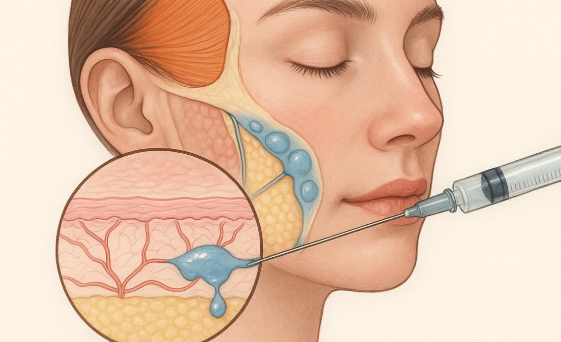
Managing superficial intradermal placement or dynamic anatomical zones demands a nuanced understanding of soft-tissue filler mechanics. In regions like the tear trough, perioral lines, or fine superficial rhytids, standard volumizing gels frequently fail. The anatomical constraints—thin skin, minimal subcutaneous fat, and high muscular activity—leave zero allowance for lumpiness, migration, or the Tyndall effect.
Predictable outcomes in these delicate zones depend less on a gel’s hyaluronic acid (HA) concentration and far more on its rheological profile. Selecting a gel based solely on duration or volume capacity often leads to visible contour irregularities. Instead, clinicians must evaluate how rheological properties—specifically $G’$ (elastic modulus), $G”$ (loss modulus), viscous flow, and cohesivity—interact with local tissue dynamics.
The Dynamic Mechanics of Hyaluronic Acid: Beyond Elastic Modulus ($G’$)
For years, dermal filler selection revolved almost entirely on elastic modulus ($G’$). While $G’$ measures a gel’s resistance to deformation and its capacity to lift under force, focusing strictly on stiffness creates a incomplete clinical picture. High-$G’$ gels excel in deep periosteal boluses where rigid support is required, but placed superficially, they remain palpably stiff and visually unnatural.
To achieve seamless blend at superficial depths, two additional properties dictate success:
- Cohesivity: The affinity of cross-linked HA molecules to stick to one another under shear stress. High cohesivity holds a gel together as a single unit, whereas low cohesivity allows the gel to disperse smoothly into adjacent intercellular spaces.
- Viscosity & Extrudability: Dictated by polymer length and cross-linking technology, low-viscosity gels flow effortlessly through fine-gauge needles (30G–32G), reducing extrusion force and minimizing micro-trauma during delicate fan or linear threading techniques.
When placing HA into the superficial dermis, high cohesivity paired with moderate-to-low $G’$ allows the material to spread evenly between collagen fibers without fragmenting into isolated nodules. This balance prevents surface unevenness while maintaining uniform hydration and soft structural support.
Tissue Integration and the Risk of Vascular/Visual Artifacts
Superficial placement introduces specific complications that do not occur in deep subcutaneous planes. The Tyndall effect—a Rayleigh scattering phenomenon where light reflects off dense, non-integrated HA particles to create a bluish discoloration—is almost always a function of mismatched gel structure and depth.
Gels with particulate properties or excessive cross-linking density often fail to integrate into the extracellular matrix (ECM) when injected superficially. They sit as distinct, particulate islands. In contrast, mono-phasic HA matrices engineered with high cohesivity and polydensified cross-linking weave directly into the dermal collagen network. This homogeneous tissue integration diffuses light naturally, eliminating the blue hue associated with poor placement.
Matching the physical properties of the implant to the surrounding tissue density dictates whether the gel acts as an internal support or an artificial obstruction.
Matching Rheology to Anatomical Indications
Evaluating dermal fillers requires balancing structural support against soft-tissue fluidity. For delicate zones, products manufactured using Dynamic Cross-Linking Technology (CPM) offer a spectrum of densities designed for varying dermal depths.
+——————+———————–+———————+——————————-+
| Indication Zone | Target Depth | Rheological Profile | Clinical Objective |
+——————+———————–+———————+——————————-+
| Fine Lines | Superficial Dermis | Low G’, High CPM | Smooth spread without lumps |
| Tear Trough | Perioridicular Sub-P. | Mod G’, Low Viscosity| Minimal water uptake, natural |
| Perioral / Lips | Mid-Dermis / Submucosa| Balanced G’/Loss Mod| Dynamic flex with movement |
+——————+———————–+———————+——————————-+
When stocking treatment rooms, purchasing teams and medical directors must weigh these physical characteristics alongside inventory versatility. Practices seeking to maintain reliable clinical supply lines can order Belotero products for clinic use through verified distribution channels to ensure authentic, temperature-controlled inventory. Reviewing full product portfolios allows clinical staff to match specific gel viscosities—from low-density superficial formulations to higher-density structural matrices—directly to patient anatomical needs.
Maintaining an optimal supply of varied viscosities ensures that clinicians never have to compromise by using a sub-optimal $G’$ gel in a fragile anatomical area. A common misstep is attempting to adapt a single versatile filler across all depths; using a mid-tier volume gel superficially in the tear trough almost guarantees late-onset fluid accumulation or palpable nodularity.
Decision Framework: Selecting the Correct Gel Viscosity
To avoid selection errors in thin-skinned or dynamic areas, clinicians can use a systematic decision process before needle insertion:
- Assess Epidermal/Dermal Thickness
- Thin / Atrophic (<1mm): Mandates a low-$G’$, highly cohesive, monophasic gel. Avoid high-swelling capacity formulations.
- Moderate/Thick (>1mm): Can accommodate moderate-$G’$ gels with higher lifting capacity.
- Evaluate Dynamic Movement
- High Movement (Perioral, Orbicularis Oculi): Requires high tissue integration and low flow resistance under shear stress. The gel must bend with facial expression without projecting outward.
- Static (Deep Sulci, Periosteal): Priority shifts toward high $G’$ and structural resistance to compression.
- Determine Hydrophilic Risk
- Regions prone to fluid retention (infraorbital area) require formulations with controlled swelling ratios to prevent post-procedural edema weeks after injection.
Common Selection Mistakes in Clinical Practice
Even experienced injectors encounter complications when rheology is overlooked. The three most prevalent miscalculations include:
- Over-relying on Concentration over Cross-linking: High HA concentration (e.g., 25 mg/mL) does not automatically mean high lifting power or long duration. The balance of cross-linking ratio and molecular weight determines behavior in tissue, not just raw milligram count.
- Ignoring Swelling Capacity: Hyaluronic acid is inherently hydrophilic. Injecting high-swelling capacity gels near the periorbital lymphatic network frequently leads to chronic malar edema.
- Underestimating Shear Forces: In perioral applications, muscle contraction exerts continuous shear stress on the implant. Gels lacking sufficient cohesivity will break apart into small micro-spheres, migrating away from the vermilion border.
Practical Considerations for Product Integration
Integrating specialized HA gels into daily practice requires consistent protocols. Storage condition monitoring, sterile prep, and precise delivery tools (such as 30G–32G extra-thin wall needles or blunt-tip 27G microcannulas) play an equal role in final outcomes. While correct gel choice is critical, controlled depth of administration remains the ultimate determinant of aesthetic success.
By aligning anatomical requirements with precise rheological characteristics—cohesivity, viscosity, and elastic modulus—clinicians consistently achieve smooth tissue integration, low complication rates, and natural dynamic outcomes in the face’s most challenging zones.