
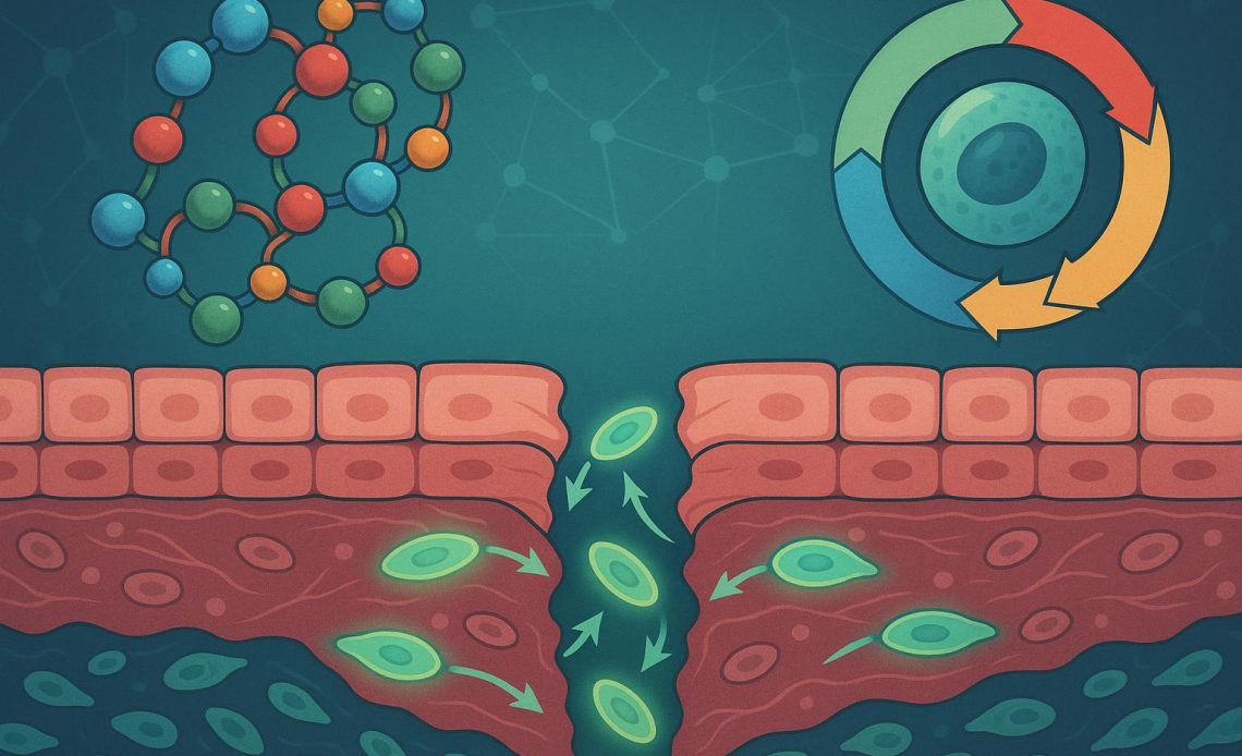
Endogenous peptides act as critical signaling intermediaries, coordinating localized cell communication, gene expression, and homeostatic maintenance. Within preclinical research, specific short-chain peptide sequences—frequently categorized as bioregulators—have drawn interest due to their capacity to modulate targeted physiological processes at the genomic and epigenetic level. Investigating these molecules provides key insights into tissue regeneration kinetics, cellular senescence, and the signaling pathways governing cell cycle progression.
Structural Characteristics and Mechanisms of Bioregulator Peptides
Unlike larger protein structures or high-molecular-weight biologicals, bioregulator peptides typically consist of short amino acid sequences, often ranging from two to four residues. This lower molecular weight facilitates specific nuclear translocation, allowing these short sequences to interact directly with targeted histone proteins and DNA promoter regions.
In laboratory settings, research demonstrates that these micro-peptides bind to the major and minor grooves of DNA in a sequence-specific manner. This interaction can alter chromatin structure, influencing the accessibility of transcription factors without altering the primary DNA sequence itself.
- Transcriptional Modulation: Selective upregulation or suppression of specific structural and functional proteins.
- Epigenetic Regulation: Influence over DNA methylation patterns and histone acetylation states during cellular aging models.
- Telomerase Activation: Variable modulation of telomerase activity in senescence models, affecting long-term cell passage limits in culture.
The sequence-specific binding mechanism allows these short oligopeptides to operate as direct modulators of chromatin accessibility. When a bi- or tetrapeptide enters the cell nucleus, its specific side-chain charges align with matching base-pair sequences across target genes. This physical binding induces localized conformational shifts in the DNA-histone complex, essentially unpacking heterochromatin into euchromatin and permitting RNA polymerase access.
Signaling Pathways in Preclinical Tissue Repair
When evaluating tissue repair dynamics in vitro, researchers focus on how short-chain sequences interface with standard repair cascades. Following localized tissue strain or cellular damage, signal transduction relies on a balance between pro-inflammatory cytokine expression and matrix synthesis factors.
In culture models examining mesenchymal and epithelial regeneration, specific peptide sequences exhibit modulating effects on Transforming Growth Factor-beta (TGF-β) signaling and Nuclear Factor kappa B (NF-κB) activation. By tempering overactive inflammatory cascades, these molecules allow cell cultures to maintain structural integrity and proceed through standard repair phases.
| Receptor Target / Pathway | Primary In Vitro Observation | Research Focus Area |
|---|---|---|
| TGF-β / Smad Pathway | Modulation of extracellular matrix (ECM) deposition rates | Fibrotic response control, collagen synthesis models |
| NF-κB Cascade | Suppression of nuclear translocation of inflammatory subunit p65 | Cellular stress response, inflammatory signaling |
| MAPK / ERK Pathway | Upregulation of baseline cell proliferation indicators | Wound closure rate kinetics, epithelial migration |
| Cyclin/CDK Complexes | Regulation of G1-to-S phase transition checkpoints | Cell cycle arrest mechanisms, senescence delay |
Wound healing models frequently utilize scratch assays in epithelial monolayer cultures to track cellular migration rates. When short-chain peptides are added to these assays, researchers often observe a reduction in time to wound closure. This accelerated closure is generally mediated through upregulated Focal Adhesion Kinase (FAK) signaling alongside increased expression of cell-adhesion molecules like E-cadherin, indicating enhanced cell motility rather than uncontrolled hyper-proliferation.
Material Selection and Experimental Standards in Peptide Research
When designing protocols to analyze epigenetic modulation or tissue repair kinetics, maintaining high assay reproducibility requires strict chemical purity and analytical validation of test compounds. Minor variations in sequence fidelity, counter-ion content, or synthetic residual impurities can alter binding affinity curves or induce non-specific cellular stress responses.
Laboratory assays frequently evaluate specific sequence classes to map out binding affinities, degradation rates in physiological buffers, and downstream expression profiles. Laboratories sourced for analytical work rely on standardized vendors to shop bioregulator peptides for laboratory research, ensuring access to validated purity profiles verified via HPLC and mass spectrometry. Utilizing characterized sequences allows investigators to systematically benchmark observed biological activity against baseline parameters without confounding variables from chemical degradation.
Following compound selection, researchers analyze expression changes using high-throughput RNA sequencing (RNA-seq) or targeted quantitative PCR (qPCR) panels. These assays verify whether observed phenotypic shifts correlate directly with predicted transcriptional alterations. High-purity peptides ensure that observed transcriptional responses stem specifically from peptide-DNA interactions rather than batch contaminants like residual trifluoroacetic acid (TFA) salts or truncated peptide fragments.
Cell Cycle Control and Senescence Research Models
Understanding how cellular populations transition into senescence remains a major objective in cell biology. Senescent cells exhibit altered secretion profiles—often referred to as the senescence-associated secretory phenotype (SASP)—which can degrade adjacent extracellular matrix structures and disrupt surrounding cell function in co-culture systems.
Preclinical work with short-chain peptides explores their ability to influence cell cycle arrest machinery, particularly through pathways mediated by p16INK4a and p21CIP1/WAF1.
In models of accelerated senescence, specific oligopeptides demonstrate an ability to suppress SASP component release—such as Interleukin-6 (IL-6) and Matrix Metalloproteinase-3 (MMP-3)—while preserving baseline proliferative capacity across extended cell passage numbers. By maintaining CDK (cyclin-dependent kinase) activity within normal physiological ranges, these compounds provide a valuable experimental lens through which to study the prevention of premature cell cycle arrest.
Furthermore, cellular aging assays regularly measure beta-galactosidase activity as a primary biomarker of senescent burden. When senescent cultures are exposed to target peptide sequences, researchers frequently record a measurable decrease in senescence-associated beta-galactosidase (SA-β-gal) staining, pointing to a potential restoration of youthful transcriptional activity in primary cell lines.
Framework for Laboratory Evaluation of Peptide Activity
To ensure robust data collection when evaluating short-chain sequences in vitro, research groups typically follow a structured assessment framework:
- Analytical Verification: Confirm purity using High-Performance Liquid Chromatography (HPLC) and confirm molecular weight via Mass Spectrometry (MS).
- Solubility & Stability Testing: Determine sequence stability in target physiological buffers (e.g., PBS, cell culture media) across standard temperature ranges over designated timepoints.
- Cytotoxicity & Proliferation Assays: Establish baseline viability parameters using standard colorimetric assays (e.g., MTT, XTT) prior to running functional experiments.
- Target Gene Expression Analysis: Map transcriptional shifts using qPCR or transcriptomic profiling to verify sequence-specific mechanism of action.
- Phenotypic / Functional Quantification: Measure downstream outcomes such as cell migration rates (scratch assays), protein synthesis levels, or senescence marker expression.
Evaluating these parameters systematically prevents false-positive conclusions driven by altered media pH, osmolarity changes, or peptide aggregation. Stability profiling is particularly essential, as short peptides are prone to rapid cleavage by endogenous peptidases present in fetal bovine serum (FBS) added to standard culture media.
Methodological Limitations and Future Scope
While in vitro data regarding bioregulator peptides provides clear insights into nuclear binding and transcriptional control, significant analytical challenges remain. Peptides inherently face enzymatic degradation in biological fluids, requiring careful optimization of experimental conditions, such as short incubation windows or specialized delivery vehicles (e.g., lipid nanoparticles or specialized culture matrixes).
Furthermore, observing a gene expression change in isolated cell lines does not automatically map to multi-organ system interactions. Future preclinical efforts continue to focus on mapping out the precise biophysical interactions between short sequences and specific histone markers, establishing more detailed structural models of micro-peptide chromatin regulation.
Advanced spectroscopic techniques, including nuclear magnetic resonance (NMR) spectroscopy and X-ray crystallography, are increasingly utilized to map the exact binding interfaces between di- and tetrapeptides and DNA major grooves. As computational modeling tools mature, predictive algorithms will likely accelerate the discovery of novel short-chain sequences tailored for specific cellular target pathways.