
The Ken Mwatha is a board-certified emergency medicine physician who serves as an attending physician in the Department of Emergency Medicine at St. Agnes Hospital in Baltimore, Maryland. With more than a decade of experience in high-acuity emergency care, he delivers life-saving treatment for severe trauma and illness while also overseeing operational needs such as patient flow and rapid evaluation. His background includes earlier work as an attending physician at Harbor Hospital, where he managed emergent and non-emergent presentations and supported timely procedural intervention in trauma and critical-care settings. Dr. Mwatha has also contributed to emergency medicine education and research, including co-authoring material for the Johns Hopkins Emergency Medicine Intern Guidebook and participating in studies involving diagnostic imaging, inflammatory bowel disease, and HIV-related research. These roles align closely with the clinical realities discussed in bedside ultrasound use when time-sensitive decisions must be made during emergency evaluation.
How Emergency Ultrasound Helps Avoid Delays in Critical Care
Emergency departments move quickly, but delays occur when teams need answers from imaging that happens outside the room. When clinical decisions depend on late-arriving information, treatment momentum slows. Emergency physicians use bedside ultrasound to obtain focused answers during evaluation and help shorten that gap, while ensuring scans do not delay urgent treatment.
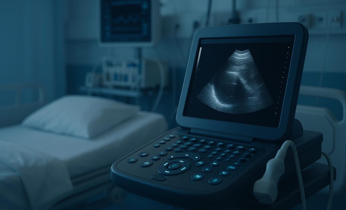
Bedside ultrasound, also called point-of-care ultrasound (POCUS), is a portable imaging tool that uses sound waves to generate real-time internal images. Emergency physicians perform the exam themselves rather than sending patients to radiology. The purpose is not a comprehensive diagnosis but a rapid clarification of specific questions, such as looking for fluid in the lungs or abdomen. Studies in emergency care show that bedside ultrasound can shorten time to diagnosis and treatment in some high-risk scenarios. However, its impact depends on the condition, setting, and the team’s training.
Consider a patient with early pregnancy symptoms and severe abdominal pain. A focused bedside scan can quickly assess for warning signs of an ectopic pregnancy, a pregnancy growing outside the uterus. When the scan raises concern, the emergency team can expedite formal imaging and specialist evaluation rather than waiting for a delayed first look.
In surgical abdominal pain, studies examine whether an emergency physician–performed ultrasound can reduce time in the department. In suspected appendicitis, for example, one study found a shorter emergency department length of stay when emergency physicians could diagnose appendicitis using bedside ultrasound alone. When ultrasound did not provide sufficient clarity, clinicians still used additional imaging, such as a computed tomography (CT) scan, to confirm the diagnosis and guide next steps.
Training focuses on consistent use of a limited set of focused views for common high-risk presentations. Practical barriers still shape whether ultrasound reduces delays, including tight time constraints, limited training for bedside staff, and the need for clear protocols.
A standard or incomplete scan does not exclude disease, and ultrasound does not replace other testing or imaging when the case requires it. Most importantly, emergency references and outcome studies emphasize that clinicians should not prioritize scanning over life-saving stabilization or other time-critical interventions. When teams use ultrasound at the wrong moment, especially before key interventions in unstable patients, it can add delay instead of removing it.
Ultrasound most effectively reduces delays when clinicians use it in a specific way. The treating emergency clinician asks one focused question, scans. It uses the result to choose the following action: treat now, consult now, observe with re-checks, or proceed to formal imaging. Ultrasound findings support decisions, but they do not replace clinical monitoring, labs, or radiology when the case requires them.
As ultrasound becomes more common in emergency medicine training, its role continues to evolve. Hospitals increasingly focus on clear rules for when to scan, who should scan, and how to record results so others can rely on them. The goal is not to treat ultrasound as the first test in every case, but to ensure the first decisions do not wait on information that the bedside team can safely obtain sooner.
Departments get the most value from bedside ultrasound when they treat it as a timed decision step rather than an extra task. Teams can define which high-risk presentations justify an early focused scan and use the same short note each time to record the clinical question and the key finding. Clinicians also maintain a hard rule: scanning never delays stabilization. With those guardrails in place, bedside ultrasound can speed escalation when it answers a narrow question early.
About Ken Mwatha
A board-certified emergency physician, Dr. Mwatha provides high-acuity care as an attending physician in the Department of Emergency Medicine at St. Agnes Hospital in Baltimore. He previously served as an attending physician at Harbor Hospital, treating emergency conditions and supporting department operations. His research and education experience includes contributing to the Johns Hopkins Emergency Medicine Intern Guidebook and participating in projects involving diagnostic imaging, inflammatory bowel disease, and HIV. Outside of medicine, he follows rugby and competed on the University of Wyoming’s rugby team.